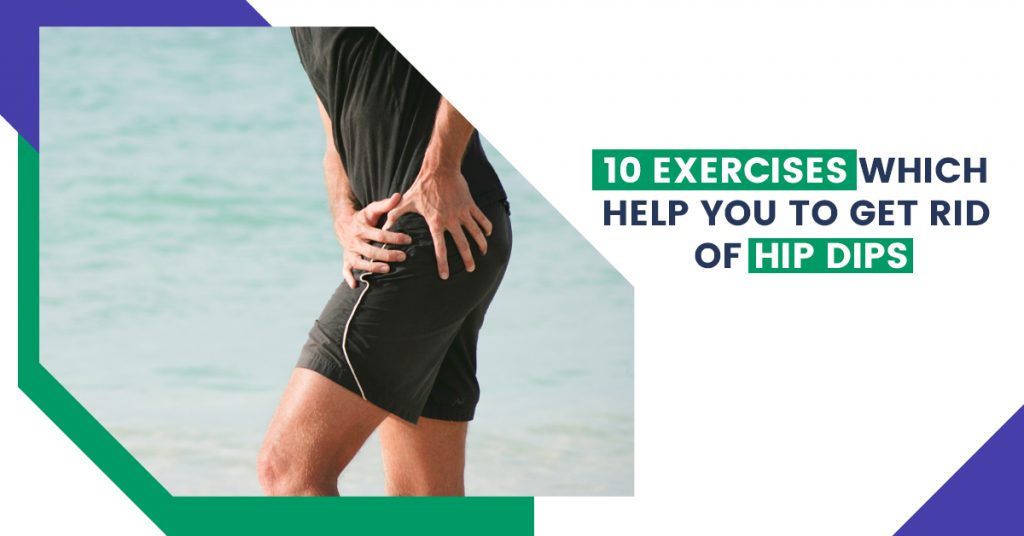
10 Exercises Which Help You to Get Rid of Hip Dips
In order to maintain good health, our bodies require regular exercise. The idea of exercising is not to spend hours in the gym or to do heavy workouts. Exercises that tone up your muscles and remove marks can also help you achieve a healthy and attractive body.
The hips play a crucial role in giving the body its ideal structure. Therefore, everyone aspires to have rounded, well-structured hips. Sometimes, however, hip dips occur in the body due to an excess of fat or bone structure. Let’s study hip dips today and find out which exercises you should do in order to eliminate them.
What are hip dips?
Hip dips commonly known as violin hips are a fairly typical physical characteristic of female bodies. Some ladies wish they didn’t have them, while others do.
The phrase describes a tiny depression in the hips that prevents them from having the full, rounded shape that many women desire. Even if they are not the form you want in your hips, they are a typical feature of the body structure and are not brought on by being overweight.
Hip dips vary greatly from person to person and can be extremely mild or very apparent. With diet and exercise, some people can reduce their appearance while others cannot. It’s also crucial to understand that hip dips and love handles are two different things.
What causes hip dips?
Genetics determines the skeletal structure of your hips, which results in hip dips.
Your hip dips will be visible based on the following factors:
- how wide are your hips are
- Having a large greater trochanter (the top of your femur)
- The distances between your greater trochanter, hip socket, and ilium (a portion of your pelvis).
- Your femoral neck’s length
- Your fat distribution
- Your muscle mass
Your hips and buttocks’ form and the presence of hip dips are both significantly influenced by these elements.
Hip dips happen where the skin is tied, or linked, to the trochanter, the deeper portion of your thigh bone. It is more obvious in some people because of how much and how evenly their bodies are made up of muscle and fat.
The width of your hips, the form of your pelvis, the distribution of your body fat, and other factors might make hip dips more or less noticeable. Additionally, it’ll be easier to see if you’re wearing tight combat attire like leggings or slim pants.
There are workouts that can assist in keeping hips under control, even though we can’t entirely get rid of them.
You can Read Also: Hyperspermia: Causes, Symptoms, Fertility & Treatment
10 Best Exercises Which Help You to Get Rid of Hip Dips
The simplest technique to lessen hip dips’ appearance is to specifically target them with exercise moves. Exercises for the hip dip don’t have to be difficult. Following this routine will help you get rid of hip dips because consistency is important.
You don’t even need a gym membership or expensive equipment for these hip dip workouts. Simply perform 5-7 of these exercises weekly to see the problem go.
1. Side hip openers (fire hydrants)
These exercises concentrate on your side buttocks, hips, and outer thighs.
- Begin on all fours, knees must be directly below hips, and hands should be just below shoulders.
- While raising one leg, bend the other leg at a 90-degree angle while exhaling. Keep your knee bent at all times.
- As you carefully lower your leg back down, take a breath. Before you lift it again, keep your knee from touching the ground.
- Repeat this motion 15 times. As you lower your leg on the final rep, pulse it ten times before lowering it.
2. Squats
Your butt, thighs, hips, and legs are all strengthened by squats.
- While standing, keep your feet hip-width apart.
- Breathe in and tighten your abdominal muscles as you slowly lower your booty as if you’re going to sit in a chair that doesn’t exist.
- Engage your core and keep your weight on your heels.
- Exhale as you stand up by driving your hips forward and pushing your feet into the ground.
- Do repetition 10–12 times.
3. Hip Abduction
Exercises for hip abduction can help the hips become more flexible and stronger.
- Place your top arm in front of your chest while lying on your side to support your upper body.
- Raise your top leg toward the ceiling while maintaining the most rigid and engaged upper body and core you can.
- Lower back down – with control
- Do repetition.
4. Glute Rainbows
It helps to increase the stability and mobility of the hip joint.
- On your mat, get down on all fours. Straighten your left leg behind you as you lift it.
- Sweep it behind and across your right leg while maintaining a level posture and an arcing motion.
- Then, with your left hip, sweep it past your beginning point to a lateral position.
- Repeat with the opposite leg, then head back to the centre.
5. Clamshell
The clamshell exercise can help your medial glutes become stronger, giving your hips more strength and stability.
- You can begin this exercise by lying on your side with your head resting on the arm that is on the floor.
- Your hips should be at a 45-degree angle, and your knees should be at a 90-degree angle.
- The knee should be pulled away from your centre while keeping your feet together.
- Pause and tense your glutes and abdominals as you near the peak. returning to the ground.
- Do repetition.
6. Side Lunge
Side lunges are good for strengthening your quads, glutes, and other lower body muscles.
- As you perform this exercise, place your feet together and stand at the top of your mat.
- Lunge laterally with your butt pushed behind you as you contract your core muscles.
- You should continue to apply pressure with your heel while lunging.
- Do repetition on both sides.
You can Read Also: Malaria: Causes, Symptoms, Diagnosis, and Treatment
7. Curtsy Lunge
The curtsy lunge is a fantastic exercise for developing lower body stability and strength.
- You can start by lunging backwards while standing with your feet hip-width apart.
- Cross your lunging leg across to the other side to generate a deep curtsy.
- Push through the heel of the front foot to return to the starting position.
- With the other leg, repeat the motion.
8. Glute Bridges
Glute bridges are the best exercise to do if you want to have strong, toned buttocks and fewer hip dips.
- To begin, raise your heels to the mat while lying on your back.
- Ensure that your knees point upward and your heels are a few inches away from your buttocks.
- Toes should be pointed outward and your feet should be spaced slightly apart from one another.
- Keep your knees pushed outward while working your side glutes.
- Raise your pelvis off the floor, allow it to rest for a moment, and then lower it again.
9. Step-Downs
The hips, hamstrings, and quads are worked during step-downs. They can also help stabilize the knees.
- Step up onto a strong, reasonably low seat, step, or stool.
- As you take a slow, one-footed step down, engage your glutes and core.
- Tap the ground with the lower foot.
- Return it slowly to the stool’s starting position.
- Do 10 repetitions on each side.
10. Donkey Kicks
Your butt can be toned, lifted, and strengthened with leg kickbacks.
- Start in a tabletop position while on all fours. Make sure your hips are squarely over your knees and your shoulders are over your hands.
- Once your quad is parallel to the floor, lift your knee, keeping it bent.
- Go back to the beginning place.
- Do 15 repetitions on each side.
10 Exercises Which Help You to Get Rid of Hip Dips Read More »