Introduction
The knowledge of the standard range of eye pressure is very important with respect to eye care, as well as a measure of prevention for serious diseases like glaucoma. In this guide, we’ll discuss eye pressure, intraocular pressure (IOP), the range of normal eye pressure range levels, the signs of high eye pressure , the methods used for measuring eye pressure, and the options available for you in case of abnormal IOP.
What Is Eye Pressure?
Eyeball eye pressure is the term that comes to mind when talking about the liquid pressure in the eyeball that not only keeps its shape but also functions properly. The pressure is due to a liquid called aqueous humour, which is being produced inside the eyeball and drained through tiny ducts all the time. The eye pressure is said to be normal when the amount of the produced and drained fluid is equal. The pressure could either be high or low when this balance is disrupted, either by too much liquid being produced or too little being drained, and this could lead to impaired vision and poor health of the eye.
What Is IOP?
Intraocular Pressure (IOP) is the medical term for the measurement of eye pressure. It is measured in millimetres of mercury (mmHg), just like blood pressure is. It is very important to keep the IOP within normal limits because that is one of the ways to ensure that the eye maintains its roundness and that the optic nerve, which carries the visual messages to the brain, stays healthy. Abnormal IOP levels, whether they are high or low, can lead to the development of diseases such as glaucoma or ocular hypotony, which in turn can result in painless but very hard-to-treat conditions.
You can read also:- कीमोथेरेपी: प्रकार और किस स्टेज में कितनी प्रभावी रहती हैं?
What Is the Normal Eye Pressure Range?
The standard eye pressure is more commonly expressed as a range than a single value. Here are the main points:
- Healthy eye pressure levels range: Normally between 10 and 21 millimetres of mercury (mmHg).
- Some references limit it to approximately 10 to 20 mmHg.
- The mean IOP in most people is approximately 15 to 16 mmHg.
- Individual variability is possible: a pressure of, for instance, 22 mmHg may not always indicate a disease in some individuals, whereas others may suffer from damage even at pressures that fall within the “normal” range.
- Ocular hypotony or low eye pressure is not very common, and it only happens in very few cases when the pressure drops significantly; some define < 7 mmHg as hypotony.
What Are Symptoms of High Eye Pressure?
Often, high eye pressure in the beginning stages is either completely symptomless or very subtly manifested, which is precisely the reason regular eye examinations are essential. The following is a list of the most important signs and symptoms:
- Silent progression: A large number of people with high IOP or even the first stages of optic nerve damage are not aware of anything troubling and do not even notice any changes in their vision.
- Visual disturbances: In a more advanced stage, the individual may become aware of the loss of peripheral (side) vision, tunnel vision, or trouble seeing in low-light conditions.
- Eye pain or headache: These are rare except when the pressure increases suddenly (e.g., in acute angle-closure glaucoma).
- Halos around lights or blurry vision: These can be observed during acute pressure spikes.
- Eye redness or heaviness: Those symptoms may be present along with other signs in acute cases.
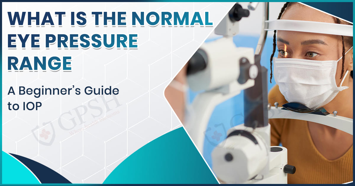
How Is Eye Pressure Measured?
Measuring intraocular pressure is an easy process, but still, there are significant aspects to pay attention to:
- Tonometry is the collective name for all the tests that check the IOP.
- The Goldmann Applanation Tonometry (GAT) is the highest standard, where a little device flattens a part of the cornea, and the force used is then transformed to pressure.
- Other techniques are non-contact “air puff” tonometry, rebound tonometry, and portable devices; usually, their accuracy is a bit lower, as these methods are often used for screening.
- The correctness of the measurement can be affected by the properties of the cornea, such as its thickness. IOP might be less or more than the real value, according to thinner or thicker corneas.
- Since IOP can change throughout the day (higher in the morning, lower afterwards), it is not uncommon that more than one measurement is taken to obtain a reliable baseline.
You can read also:- Water-Borne Diseases: Treatment, Types, Causes, Symptoms, and Prevention
Signs of High Eye Pressure
There are particular signs which will be detected by your eye doctor apart from the pressure number that include the following:
- Optic nerve deterioration indicators: The examination or imaging will show the optic nerve head changes (e.g., cupping).
- Decreased visual field: particularly, a decreased peripheral vision can be determined from visual field testing.
- High pressures: Any reading of IOP that exceeds normal amounts will be a concern.
- Presence of risk factors: Family history of glaucoma, over 40 years of age, high amount of nearsightedness, taking steroids, and history of eye trauma.
- Low IOP (hypotony): Very low IOP (for example, < 7 mmHg) may also lead to similar problems, such as the death of cells in the structure or alterations in the cornea.
Why Is It Important to Maintain Healthy Eye Pressure Levels?
The very first condition for the protection of the optic nerve and the preservation of the eyesight is the maintenance of normal intraocular pressure. Changes in eye pressure, whether too high or too low, can cause the following problems:
- Glaucoma risk: High IOP is considered the main risk factor for glaucoma, although it does not necessarily mean that all individuals with high IOP will develop glaucoma, and vice versa, some people with normal IOP may still have the disease.
- Optic nerve damage: The nerve fibers in the retina may become irreversibly lost due to prolonged elevated pressure that can either compress or damage the optic nerve.
- Structural eye issues: Hypotony, or low pressure in the eye, may cause the eye to lose its shape, leading to corneal edema, retinal detachment, and other complications.
Conclusion
The typical eye pressure of a person is mostly within the range of 10 to 21 mmHg, where the majority of healthy people have their eye pressure measured around 15 to 16 mmHg. Still, “normal” is a somewhat subjective word, and what is safe for one individual might not be so for another. A high or a low IOP can endanger your vision.
If you are not sure about the intraocular pressure of your eyes or if you are at risk for glaucoma, it is good to consult an eye care specialist. Shekhawati Hospital’s Glaucoma Department can help with a more individualized strategy for dealing with these intraocular pressure problems for those patients who need treatment or evaluation for glaucoma, which includes measuring, diagnosing, and managing intraocular pressure.
FAQs
Q1. Can someone still be diagnosed with glaucoma even if their eye pressure is normal?
Certainly, that is the case. Some specific people have an IOP within the normal clinical range, but have evidence of damage to the optic nerve and a visual field defect; that would be called normal-tension glaucoma.
Q2. How frequently should I have my intraocular pressure checked?
A thorough eye examination, including IOP measurement every 1–2 years, is recommended for those aged over 40 or who have risk factors (family history, high myopia, steroid use). Higher-risk individuals may need even more frequent checks.
Q3. What actions can I take to prevent my eye pressure from rising above the normal range?
Regular check-ups and eye tests, following the advice given by the eye doctor, not using steroids heavily without supervision, controlling systemic diseases like high blood pressure or diabetes, and adopting a healthy lifestyle (physical activity, balanced diet, no smoking) that contributes to the overall health of the eyes.
Q4. What will happen if my IOP is marginally above normal, but no damage to the optic nerve is observed?
Your eye doctor might label it ocular hypertension and pay even closer attention to it. They may recommend that you begin treatment for lower IOP or continue to monitor your IOP until there are further signs.

