What Are the First Signs of Chickenpox? Symptoms, Causes, Treatment & Prevention
Introduction
Chickenpox is a very infectious virus disease that mainly children and occasionally unvaccinated or previously infected adults suffer from. The varicella-zoster virus is responsible for this skin disease and it is characterized by a burning itch and red spots which are scattered all over the body.
Early detection of the first signs of chickenpox can assist in the control of symptoms and hence the transmission of the virus to other people can be reduced. The present article is aimed at shedding light on the symptoms, causes, treatment options, and preventive measures in order to give a complete picture of chickenpox.
What Is Chickenpox?
Chickenpox, also known as varicella, is an epidemic illness caused by the varicella-zoster virus. Transmission mainly occurs through a person having direct contact with an infected individual or by way of respiratory droplets generated from coughing or sneezing. The disease typically manifests as an itchy rash made up of small, fluid-filled vesicles that ultimately dry up and turn into scabs.
Chickenpox is non-complicated in the vast majority of scenarios and simply goes away without any medical intervention. However, it might be severe in certain groups of people such as newborns, pregnant women, or patients with a compromised immunity. After one has healed, the virus remains in hiding inside the body and can later come back as shingles.
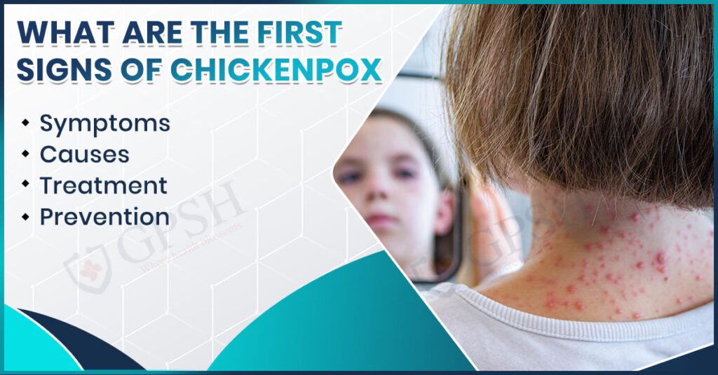
Symptoms & First Signs of Chickenpox
Typically, the initial signs of chickenpox manifest after a span of 10 to 21 days post contact with the virus. A patient would experience a mild, flu-like condition prior to the appearance of the rash. The following is a list of symptoms that you need to observe:
- Early Symptoms: These are the symptoms which usually come one or two days prior to the rash’s appearance.
- Fever: The mild to moderate fever is one of the first signs of chickenpox.
- Tiredness and Weakness: The immune system starts reacting to the virus, thus the body feels tired.
- Loss of Appetite: Many people have no interest in eating because of the fever or the discomfort in the body.
- Headache and Body Aches: Dull headaches and mild muscle pains are common signs occurring before the rash.
- Rash Development: The rash accompanies and goes through three stages after the first symptoms:
- Red Spots: Small, itchy red bumps first develop on the chest, face and back then spread to other areas of the body.
- Blisters (Vesicles): The duration of turning these red spots into fluid-filled blisters is a day or two.
- Crusting and Scabbing: The blisters finally break and as the infection starts to heal crusts or scabs are formed.
- Itching and Discomfort: The rash brings with it itching of the highest intensity, which can be worse at night. Scratching should be avoided to prevent both scarring and secondary infections.
You can read also:- What Is the Normal Eye Pressure Range? A Beginner’s Guide to IOP
Causes of Chickenpox
Chickenpox is the result of the varicella-zoster virus (VZV) infection, which belongs to the family of herpesviruses. The following details how it spreads and develops:
- Direct Contact: The transmission of the virus occurs via direct contact of the skin with the rash or blisters of the person infected.
- Airborne Transmission: In case the person infected coughs or sneezes, the tiny droplets containing the virus can infect others in close proximity.
- Contaminated Surfaces: This is hardly a common route of infection but it can happen that one gets the virus in the mouth or nose after touching the surface that was previously contaminated.
- Reactivation of Virus: People who have already had chickenpox, the virus can become active again later in life and shingles occur.
Treatment for Chickenpox
The majority of the instances of chickenpox heal on their own within a period of 7 to 10 days and do not need any serious medical intervention. The treatment mainly aims at alleviating the symptoms and preventing any complications. The following are the main methods:
Home Care:
- Rest and Hydration: A person should rest very well and take a lot of fluids in order to remain hydrated.
- Cool Baths: The itching and skin irritation can be relieved by taking cool or oatmeal baths.
- Loose Clothing: Soft and loose-fitting clothing is the best choice as it does not irritate the skin and reduces itching.
Medications:
- Antihistamines: Antihistamines maintain their very strong position in treating itching and giving comfort.
- Pain Relievers: Acetaminophen (paracetamol) is one of the most common medicines for managing fever and body aches. Aspirin, on the other hand, should be avoided as it is associated with Reye’s syndrome in children..
- Antiviral Drugs: Doctors may prescribe antiviral medicines like acyclovir for severe cases or those at high risk (pregnant women, adults, or immunocompromised patients).
Preventing Complications:
- Avoid Scratching: Infection and scarring will most probably occur if the area is scratched.
- Keep Nails Short: Lengthy nails will more likely cause the child to hurt himself/herself during scratching, so it is better to keep them trimmed.
You can read also:- Water-Borne Diseases: Treatment, Types, Causes, Symptoms, and Prevention
Prevention of Chickenpox
Vaccination plus good hygiene is the best prevention for chickenpox.
- Vaccination: The chickenpox Varicella vaccine usually provides long-term immunity as well as quite effective protection from illness It is generally recommended for two doses, the first dose is administered to an infant (12 to 15 months of age) and the second dose is given to a young child (4 to 6 years).
- Post-Exposure Vaccination: When there has been an exposure to a person with chickenpox and no vaccination, the vaccination may help prevent or lessen the degree of illness if received with three to five days of exposure.
- Isolation of Infected Individuals: Health authorities will suggest infected individuals to isolate until all the blisters have crusted and dried which is a recommendation to prevent the exposure of this infection to a other people.
- Hygiene: Hand washing, avoiding face contact, and disinfecting commonly used surfaces should minimize the chance of spread of this illness.
- Support Immunity: A well nourished person should, with good nutrition, regular physical activity and adequate sleep, help guide the body to proper immune responses to infections.
Conclusion
It is important to recognize the early signs of chickenpox, so that the appropriate treatment can begin and the progression of the disease can be managed. Although most cases are mild and manageable at home, it is good practice to consult your physician if the symptoms become severe or complications develop.
To get a medical diagnosis and full treatment, visit the Dermatology Department of Shekhawati Hospital, Jaipur. They are focused on treating patients with the best service on an outpatient or inpatient basis for chickenpox and other skin infections. Shekhawati Hospital has knowledgeable physicians and the latest and most up-to-date facilities/environment to provide patients with adequate management, good recovery time, and full support to prevent infections from occurring in the future.
FAQs About Chickenpox
- How long does chickenpox last?
Chickenpox generally lasts for 7 to 10 days, that is, the time taken from the first appearance of symptoms to the complete healing of scabs.
- Can adults get chickenpox?
Yes, adults who have not experienced chickenpox or received the vaccine are still able to become infected, and the severity of the infection is higher than in children.
- How do I know if it’s chickenpox or something else?
The chickenpox rash usually appears first on the face, chest, and back, then it changes from small red spots to blisters filled with fluid and, finally, to crusty scabs. When in doubt, get a doctor’s opinion for a correct diagnosis.
- Is chickenpox dangerous during pregnancy?
Yes, chickenpox during pregnancy can result in serious complications for both the mother and the baby. Pregnant women should immediately inform their gynecologist in case of exposure.
What Are the First Signs of Chickenpox? Symptoms, Causes, Treatment & Prevention Read More »