Top 10 Healthy Summer Foods You Must Include in Your Diet
When it’s hot outside, you’ll want to eat lighter meals that help keep you cool and well hydrated. This is why your body craves summer-related foods! The best part about choosing summer’s healthy foods is that in addition to helping to keep you cool, they can also give you added energy throughout the day, improve your digestion, and help you prevent dehydration. As a result, by planning a healthy summer diet, you can make a significant impact on how you feel on hot days.
What are healthy summer foods?
As you can imagine, summer’s healthy foods will help you get through the heat, stay hydrated, and replenish your energy throughout the day. Healthy summer foods are:
– High in water
– Easy to digest
– Loaded with vitamins and minerals
– Naturally cool the body
Summer foods that meet these criteria include: watermelon, cucumbers, curds/curds of curd, and coconut water. These cooling summer foods will not only feel good when you eat them, but also help you maintain balance within your body.
Why Your Body Needs Special Nutrition in Summer
During summertime sweat will remove excess amounts of fluid from your body leading you to experience dehydration; fatigue; and weakness. The summer diet is focused on the following four key principles:
- Replace fluid that has been lost.
- Keep your electrolytes balanced.
- Support your digestion.
- Prevent heat-related illness.
Eating the proper foods to prepare your body for summer will allow it to adapt to heat and will allow you to be active every day.
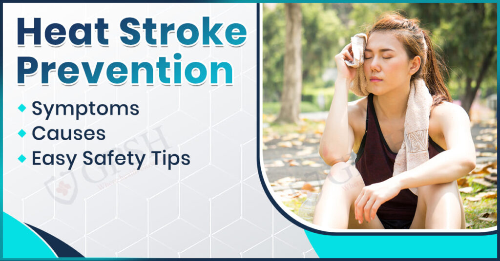
You can read can also:- Heat Stroke Prevention: Symptoms, Causes & Easy Safety Tips
Poor Summer Diet Symptoms
If your diet doesn’t match what the season requires; it is likely that you could experience the following symptoms:
- Fatigue all of the time.
- Dehydrated with dry skin.
- Frequent headaches.
- Indigestion such as gas or heartburn.
- Heat exhaustion.
All these signs signal that you are low in the nutritional needs and lack hydration.
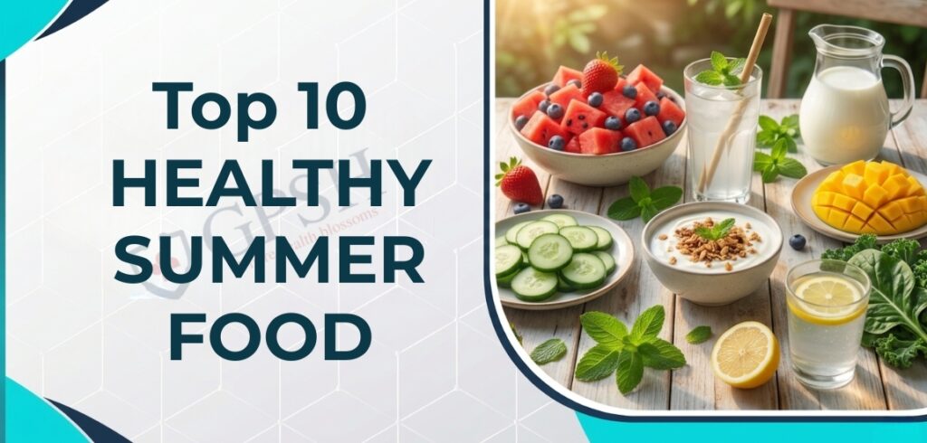
10 Healthy Summer Foods You Must Include in Your Diet
Some healthy summertime foods you should include in your diet are:
1) Watermelon
Composed of about 91% water, watermelon offers a great source of hydration and will cool you off.
2) Cucumber
Cucumbers help you cleanse toxins out of your body and keep you hydrated.
3) Coconut Water
An all-natural electrolyte drink that provides hydration and energy.
4) Curd/Yogurt
Curd contains probiotics, which promote healthy digestion and keep your intestines functioning properly.
5) Mint
Mint is a naturally cooling herb that helps stimulate digestion.
6) Lemon Water
Lemon water contains vitamin C for good health and provides a detoxifying mechanism in the body to maintain freshness.
7) Buttermilk
Buttermilk is an age-old summertime beverage that aids in healthy digestion and keeps the body cool.
8) Mangoes (consume in moderation)
Mangoes are delicious; however, eat them only occasionally as they produce a great amount of heat.
9) Leafy Vegetables
Spinach and lettuce are light on the stomach, as well as provide essential nutrients to the body.
10) Blueberries/Strawberries
Blueberries and strawberries are full of antioxidants that assist in counteracting the damages done by heat.
You can read also:- Lifestyle Diseases: Causes, Symptoms & Easy Prevention Tips
What Are the Health Benefits of Eating Seasonal Summer Foods?
Seasonal Eating has been around for centuries, and science supports it as well as tradition. Below is a list of reasons to eat seasonal foods:
- Improved Hydration: Seasonal fruits have a high water content.
- Better Digestion: Eating light foods means the digestive system works less for digestion.
- Better Immune System: Seasonal fruits are loaded with vitamins and antioxidants, which support a healthy immune system.
- Natural Cooling System: Eating seasonal fruits helps maintain a comfortable body temperature.
- More Nutrition: Freshly picked seasonal produce is higher in nutritional value than produce that has been shipped from far away.
A balanced healthy summer diet helps keep your body aligned with Mother Nature!
How to Stay Healthy and Hydrated in Summer
Good health during the summer does not just come from the foods you eat; there are other things you can do to stay healthy this summer. Here are some ideas:
- Drink 8-10 glasses of water each day.
- Eat lots of fruits and vegetables at every meal.
- Don’t skip meals.
- Eat smaller portions 6-8 times each day.
- Limit caffeine and sugary drinks.
- Take your water with you when you leave home!
Foods to Avoid During Summer
Some foods can help keep you cool in the summer, while others may feel heavy and uncomfortably hot to eat during hot months. You should probably avoid:
- Fried, greasy foods
- Spicy foods
- Excess coffee/tea
- Sodas
- Heavy desserts
- Processed/packaged foods
Conclusion
In summary, the most important thing for keeping yourself healthy, full of energy, and hydrated during the hot months is to consume a well-balanced diet suitable for the summer season. Eating the right foods can help protect your body from heat injury and promote your health.
At Shekhawati Hospital Jaipur, we are advocates of taking a preventive approach to healthcare. One of the most important things you can do for your overall health and well-being is to make better food choices based on the seasons. This summer, make smart food choices, and take good care of yourself.
FAQs
1. Which summer foods are healthy and delicious?
Ans. Examples of summer foods include garden-fresh cucumbers, coconut milk, yogurt, melons, and leafy green salad veggies.
2. What are some natural ways to hydrate?
Ans. You can hydrate your body by consuming sufficient amounts of water daily along with eating fruits, vegetables, and buttermilk.
3. Why should I avoid spicy foods in the hot weather?
Ans. Spicy foods raise your core temperature and can lead to excessive fluid loss from sweating or having stomach upset.
Top 10 Healthy Summer Foods You Must Include in Your Diet Read More »